
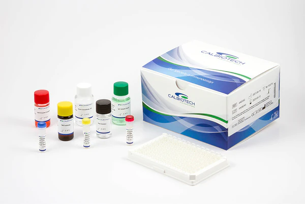
Scl-70 IgG ELISA

- 貨號 Cat. No.
- SC043G
- 規格 Size
- 96 Tests
- 品牌 Brand
- Calbiotech
Description
Scl-70 IgG ELISA
Category Name
Autoimmune Disorders
Method
ELISA
Principle
ELISA
Controls Standards
Positive, Negative and Calibrator
Sensitivity
Not available
Sample
100 µl
Runtime
50 Minutes
Wavelength
450 nm
Safety
Please refer to MSDS
Shelflife
18 Months
INTENDED USE
The Calbiotech Sm IgG ELISA Kit is intended for the detection of IgG antibody to Sm in human serum or plasma.
SUMMARY AND EXPLANATION
Systemic autoimmune disease is characterized by the presence of circulating auto-antibodies directed to a wide variety of cellular antigens. Systemic lupus erythematosis (SLE), commonly referred to as Lupus is the best known of these diseases. Other possible connective tissue diseases include mixed connective tissue disease (MCTD), Sjogren syndrome, sclerodema, and polymyositis/dermatomyositis. The majority can be diagnosed by clinical presentation and their antibody profiles to the various antigens involved, which include dsDNA, SM, RNP, SSA, SSB Scl-70, Jo1 and Histones. Therefore, immunoassays for autoantibodies are useful for diagnostic and prognostic evaluations of autoimmune disease. Scl-70 IgG antibodies react with human topoisomerase I of 100 kd molecular weight as well as its 70 kd fragment. Scl-70 antibodies are present in 20-40% of diffuse scleroderma patients and in about 20% of patients with limited scleroderma. Scl-70 antibodies are sometimes reported in classical SLE without features of scleroderma, which may explain the unexpected co-existence of marker autoantibodies for SLE and scleroderma. Some patients with silica-associated systemic sclerosis (SSc) have Scl-70 antibodies.
PRINCIPLE OF THE TEST
Diluted patient serum is added to wells coated with purified antigen. IgG specific antibody, if present, binds to the antigen. All unbound materials are washed away and the enzyme conjugate is added to bind to the antibody-antigen complex, if present. Excess enzyme conjugate is washed off and substrate is added. The plate is incubated to allow the hydrolysis of the substrate by the enzyme. The intensity of the color generated is proportional to the amount of IgG specific antibody in the sample.
