
Antibodies


WP1066 is a novel inhibitor of JAK2 and STAT3 with IC50 of 2.30 μM and 2.43 μM in HEL cells; shows activity to JAK2, STAT3, STAT5, and ERK1/2 not JAK1 and JAK3.
| Size | Price | Stock | Quantity | ||||||||||||
|---|---|---|---|---|---|---|---|---|---|---|---|---|---|---|---|
| |||||||||||||||
 Distributor Tel: +886-2-2827-1197jimmy@new.abscience.com.tw
| Description | WP1066 is a novel inhibitor of JAK2 and STAT3 with IC50 of 2.30 μM and 2.43 μM in HEL cells; shows activity to JAK2, STAT3, STAT5, and ERK1/2 not JAK1 and JAK3. | |||||
|---|---|---|---|---|---|---|
| Targets | JAK2 | STAT3 | ||||
| IC50 | 2.3 μM [1] | 2.43 μM [2] | ||||
| In vitro | WP1066 markedly inhibits the growth of HEL cells carrying the JAK2 V617F mutant isoform in a dose-dependent manner with IC20, IC50 and IC80 of 0.8, 2.3 and 3.8 μM. WP1066 at concentrations of 0.5, 1.0, 2.0, 3.0, or 4.0 μM inhibits the phosphorylation of JAK2, STAT3, STAT5, and ERK1/2 without affecting the phosphorylation of JAK1 and JAK3 in erythroid leukemia HEL cells that express the JAK2 V617F isoform. [1]WP1066 at concentrations ranging from 0.5 to 3.0 μM inhibits the proliferation of AML colony-forming cells obtained from patients and that of the AML cell lines OCIM2 and K562 in a dose-dependent manner. WP1066 at concentrations of 0.5, 1.0, 2.0, 3.0, or 4.0 μM dose-dependently decreases JAK2 and pJAK2 protein levels as well as downstream phosphorylation levels of STAT3, STAT5, and AKT in OCIM2 and K562 cells. WP1066 at concentrations of 2 μM inhibits OCIM2 cell multiplication by inducing accumulation of cells at the G0-G1 phase of the cell cycle. WP1066 at concentrations of 1, 2, or 3 μM induces apoptosis in both OCIM2 and K562 cells in a dose-dependent fashion by activating procaspase-3 and cleaving PARP. [3] WP1066 at concentrations of 5 μM prevents the phosphorylation of STAT3, and at concentrations of 2.5μM WP1066 significantly inhibits cell survival and proliferation in Caki-1 and 786-O renal cancer cells. WP1066 at concentrations of 5 μM suppresses HIF1α and HIF2α expression and VEGF production in Caki-1 and 786-O renal cancer cells. [4] | |||||
| In vivo | WP1066 orally administrated at dose of 40 mg/kg once daily for 19 days significantly inhibits the tumours growth in Caki-1 xenograft mice, with decreased immunostaining of phosphorylated STAT3 and reduced length of CD34-positive vessels. [4] | |||||
| Features | Similar to its parent compound AG490, WP1066 inhibits the phosphorylation of JAK2, but unlike AG490, WP1066 also degraded JAK2 protein. | |||||
| Cell lines | HEL cells carrying the JAK2 V617F mutant isoform, HL60, K562, Raji, PEER, CMK, TM and RHN cells |
|---|---|
| Concentrations | 0 - 6 μM |
| Incubation Time | 72 hours |
| Method | The 3, [4,5-dimethylthiazol-2-yl]-5-[3-carboxymethoxyphenyl]-2-[4-sulfophenyl]-2H-tetrazolium (MTT) assay is done using an MTT-based cell proliferation/cytotoxicity assay system. Briefly, fresh low-density peripheral blood cells and various cell lines at the logarithmic phase of their growth are washed twice in RPMI 1640 containing 10% FCS and counted in a hemocytometer. Cell viability is assessed by the trypan blue (0.1%) staining method. Equal numbers of viable cells (5 × 10⁴ per well) are incubated in a total volume of 100 μL of RPMI 1640 supplemented with 10% FCS alone or with WP1066 at increasing concentrations; the incubations are continued for up to 72 h in 96-well flat-bottomed plates at 37 °C in a humidified 5% CO2 atmosphere. Experiments for each condition are done in triplicate. After incubation, 20 μL of CellTiter96 One Solution Reagent are added to each well. The plates are then incubated for an additional 60 min at 37 °C in a humidified 5% CO2 atmosphere. Immediately after incubation, absorbance is read using a 96-well plate reader at a wavelength of 490 nm. |
| Animal Models | Caki-1 xenograft mice |
|---|---|
| Dosages | 40 mg/kg |
| Administration | Orally administrated |
| NCT Number | Recruitment | Conditions | Sponsor /Collaborators | Start Date | Phases |
|---|---|---|---|---|---|
| NCT01904123 | Not yet recruiting | Brain Cancer|Central Nervous System Neoplasms|Melanoma|Solid Tumors | M.D. Anderson Cancer Center|National Institutes of Health (NIH) | 2013-11 | Phase 1 |
| Molecular Weight (MW) | 356.22 |
|---|---|
| Formula | C17H14BrN3O |
| CAS No. | 857064-38-1 |
| Storage | 3 years -20℃Powder |
|---|---|
| 6 months-80℃in DMSO | |
| Syonnyms | N/A |
| Solubility (25°C) * | In vitro | DMSO | 72 mg/mL (202 mM) |
|---|---|---|---|
| Water | <1 mg/mL (<1 mM) | ||
| Ethanol | <1 mg/mL (<1 mM) | ||
| * <1 mg/ml means slightly soluble or insoluble. * Please note that Selleck tests the solubility of all compounds in-house, and the actual solubility may differ slightly from published values. This is normal and is due to slight batch-to-batch variations. | |||
| Chemical Name | (S,E)-3-(6-bromopyridin-2-yl)-2-cyano-N-(1-phenylethyl)acrylamide |
|---|
| Stock Solution (1ml DMSO) | 1mM | 10mM | 20mM | 30mM |
|---|---|---|---|---|
| Mass(mg) | 0.35622 | 3.5622 | 7.1244 | 10.6866 |
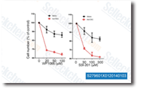 Click to enlarge | Rating | |
| Source | WP1066 purchased from Selleck | |
| Method | MTT | |
| Cell Lines | monolayer and adherent CSC | |
| Concentrations | 30, 100, 300 uM uM | |
| Incubation Time | 72 h | |
| Results | flow cytometric analysis of annexin V-stained cells demonstrated that treatment with the STAT3 inhibitor WP1066 or S3I-201 had a greater effect on the induction of apoptosis in adherent CSCs than in glioma monolayers. |
Constitutive activation of signal transducer and activator of transcription 3 (STAT3) and nuclear factor κB signaling in glioblastoma cancer stem cells regulates the Notch pathway. [Garner JM, et al. J Biol Chem 2013;288(36):26167-76]
Activation of a positive feedback loop involving IL-6 and aromatase promotes intratumoral 17β-estradiol biosynthesis in endometrial carcinoma microenvironment. [Che Q ,et al. Int J Cancer 2013;10.1002/ijc.28679]
