
Antibodies


Vandetanib (ZD6474) is a potent inhibitor of VEGFR2 with IC50 of 40 nM.
| Size | Price | Stock | Quantity | ||||||||||||||||
|---|---|---|---|---|---|---|---|---|---|---|---|---|---|---|---|---|---|---|---|
| |||||||||||||||||||
 Distributor Tel: +886-2-2827-1197jimmy@new.abscience.com.tw
| Description | Vandetanib (ZD6474) is a potent inhibitor of VEGFR2 with IC50 of 40 nM. | |||||
|---|---|---|---|---|---|---|
| Targets | VEGFR2 | |||||
| IC50 | 40 nM [1] | |||||
| In vitro | Vandetanib also inhibits VEGFR3 and EGFR with IC50 of 110 nM and 500 nM, respectively. Vandetanib is not sensitive to PDGFRβ, Flt1, Tie-2 and FGFR1 with IC50 of 1.1-3.6 μM, while almost has no activity against MEK, CDK2, c-Kit, erbB2, FAK, PDK1, Akt and IGF-1R with IC50 above 10 μM. Vandetanib inhibits VEGF-, EGF- and bFGF-stimulated HUVEC proliferation with IC50 of 60 nM, 170 nM and 800 nM, with no effect on basal endothelial cell growth. Vandetanib inhibits tumor cell growth with IC50 of 2.7 μM (A549) to 13.5 μM (Calu-6). [1]Vandetanib displays an inhibitory effect on the basal ABCG2-ATPase. Parental and ABCG2-expressing A431 cells showed similar sensitivities toward Vandetanib. Exposure to EGFR inhibitors decreases pEGFR levels in A431 cells, with Vandetanib displaying only a moderate effect. Vandetanib displays a slight but measurable effect, whereas gefitinib, pelitinib and neratinib completely inhibit ABCG2-mediated efflux of mitoxantrone from A431/ABCG2 cells, similarly to the specific ABCG2 inhibitor Ko143. [2] Vandetanib inhibits both PC3wt and PC3R cell lines with similar IC50 of 13.3 μM and 11.5 μM, respectively. [3] Vandetanib suppresses phosphorylation of VEGFR2 in HUVEC and EGFR in hepatoma cells and inhibits cell proliferation. [4]Vandetanib causes an accumulation of cells in the G0-G1 phases in GEO and OVCAR-3 cells and increases apoptosis in OVCAR-3, ZR-75-1, MCF-10A ras, and GEO cells. Vandetanib causes a dose-dependent inhibition of EGFR phosphorylation in mouse NIH-EGFR fibroblasts and human MCF-10A ras breast cancer cells, two cell lines that overexpress the human EGFR. Vandetanib treatment results in a dose-dependent inhibition of soft agar growth in seven human cell lines (breast, colon, gastric, and ovarian) with functional EGFR but lacking VEGFR2. [5] | |||||
| In vivo | Vandetanib (2.5 mg/kg, i.v.), reverses a VEGF-induced hypotension by 63% but does not significantly affect a bFGF-induced hypotension. Vandetanib (100 mg/kg) inhibits the tumor-induced blood vessel formation by 79%. Vandetanib (12.5-100 mg/kg, orally) shows great tumor growth inhibition in human tumor xenografts including Calu-6, PC-3, MDA-MA-231, SKOV-3, SW620, A549, A431, B16-F10(AP3) and Lewis Lung, with little effects on body weight. [1] In PC3wt xenografts, administration of Vandetanib alone exerts paradoxical tumor growth stimulating effects. In PC3R xenografts, the low dose of Vandetanib (25 mg/kg) has no significant effect relative to control, whereas the high dose (50 mg/kg) significantly inhibits tumor growth compared with control. In contrast, the high-dose combination reveals a significant negative interaction between Vandetanib 50 mg/kg and docetaxel 30 mg/kg in PC3R cells. [3] In tumor-bearing mice, Vandetanib suppresses phosphorylation of VEGFR2 and EGFR in tumor tissues, significantly decreases tumor vessel density, enhances tumor cell apoptosis, suppresses tumor growth, improves survival, reduces number of intrahepatic metastases, and up-regulates VEGF, TGF-alpha and EGF in tumor tissues. Treatment with Vandetanib is not associated with serious adverse events, including ALT abnormality, bone marrow suppression or body weight loss. [4]Vandetanib treatment of nude mice bearing palpable GEO colon cancer xenografts (which are sensitive to inhibition of EGFR signaling) induces dose-dependent tumor growth inhibition. [5] | |||||
| Features | ||||||
| Kinase inhibition | Vandetanib is incubated with enzyme, 10 mM MnCl2, and 2 μM ATP in 96-well plates coated with a poly(Glu, Ala, Tyr) 6:3:1 random copolymer substrate. Phosphorylated tyrosine is then detected by sequential incubation with a mouse IgG anti-phosphotyrosine 4G10 antibody, a horseradish peroxidase-linked sheep antimouse immunoglobulin antibody, and 2,2′-azino-bis(3-ethylbenzthiazoline-6-sulfonic acid). This methodology is adapted to examine selectivity versus tyrosine kinases associated with EGFR, PDGFRβ, Tie-2, FGFR1, c-kit, erbB2, IGF-1R, and FAK. All enzyme assays (tyrosine or serine-threonine) used appropriate ATP concentrations at or just below the respective Km (0.2–14 μM). Selectivity versus serine-threonine kinases (CDK2, AKT, and PDK1) is examined using a relevant scintillation proximity-assay (SPA) in 96-well plates. CDK2 assays contained 10 mM MnCl2, 4.5 μM ATP, 0.15 μCi of [γ-33 P]ATP/reaction, 50 mM HEPES (pH 7.5), 1 mM DTT, 0.1 mM sodium orthovanadate, 0.1 mM sodium fluoride, 10 mM sodium glycerophosphate, 1 mg/mL BSA fraction V, and a retinoblastoma substrate (part of the retinoblastoma gene, 792–928, expressed in a glutathione S-transferase expression system; 0.22 μM final concentration). Reactions are allowed to proceed at room temperature for 60 minutes before quenching for 2 hours with 150 μL of a solution containing EDTA (62 mM final concentration), 3 μg of a rabbit immunoglobulin anti-glutathione S-transferase antibody and protein A SPA-polyvinyltoluene beads (0.8 mg/reaction). Plates are then sealed, centrifuged (1200× g for 5 minutes), and counted on a Microplate scintillation counter for 30 seconds. |
|---|
| Cell lines | Calu-6, PC-3, MDA-MA-231, SKOV-3, SW620, A549, A431, B16-F10(AP3) and Lewis Lung cells |
|---|---|
| Concentrations | 0.1–100 μM |
| Incubation Time | 72 hours |
| Method | Tumor cells are plated in their respective media at predetermined densities that are known to enable logarithmic cell growth during the period of assay (PC-3, 500 cells/well; all others, 1000 cells/well). Plates are incubated for 24 hours (37 °C with CO2) before the addition of Vandetanib (0.1–100 μM) or vehicle (0.1% DMSO in medium). Plates are reincubated for an additional 72 hours before assessing cell proliferation by [3H]thymidine incorporation by a beta counter. |
| Animal Models | Female athymic (nu/nu genotype) Swiss mice with PC-3, Calu-6, SKOV-3, and MDA-MB-231 tumors |
|---|---|
| Dosages | 12.5 mg/kg/day, 25 mg/kg/day, 50 mg/kg/day, or 100 mg/kg/day |
| Administration | Oral administration |
| Solubility | 30% propylene glycol, 5% Tween 80, 65% D5W, 30 mg/mL |
| NCT Number | Recruitment | Conditions | Sponsor /Collaborators | Start Date | Phases |
|---|---|---|---|---|---|
| NCT01823068 | Recruiting | Non Small Cell Lung Cancer | Seoul National University Hospital|AstraZeneca | 2013-08 | Phase 2 |
| NCT01876784 | Recruiting | Differentiated Thyroid Cancer | AstraZeneca | 2013-09 | Phase 3 |
| NCT02015065 | Recruiting | GIST | National Institutes of Health Clinical Center (CC)|National Cancer Institute (NCI) | 2013-09 | Phase 2 |
| NCT01945762 | Not yet recruiting | Symptomatic, Aggressive, Sporadic, Unresectable, Locally|Advanced/Metastatic Medullary Thyroid Cancer (MTC) | AstraZeneca|Worldwide Clinical Trials | 2013-10 | |
| NCT01941849 | Not yet recruiting | Phaeochromocytoma|Paraganglioma | University College, London|Cancer Research UK|AstraZeneca | 2014-01 | Phase 1 |
Download Vandetanib (ZD6474) SDF
| Molecular Weight (MW) | 475.35 |
|---|---|
| Formula | C22H24BrFN4O2 |
| CAS No. | 443913-73-3 |
| Storage | 3 years -20℃Powder |
|---|---|
| 6 months-80℃in DMSO | |
| Syonnyms | ZD6474 |
| Solubility (25°C) * | In vitro | DMSO | 4 mg/mL (8 mM) |
|---|---|---|---|
| Water | <1 mg/mL (<1 mM) | ||
| Ethanol | <1 mg/mL (<1 mM) | ||
| In vivo | 30% propylene glycol, 5% Tween 80, 65% D5W, | 30 mg/mL | |
| * <1 mg/ml means slightly soluble or insoluble. * Please note that Selleck tests the solubility of all compounds in-house, and the actual solubility may differ slightly from published values. This is normal and is due to slight batch-to-batch variations. | |||
| Chemical Name | N-(4-bromo-2-fluorophenyl)-6-methoxy-7-((1-methylpiperidin-4-yl)methoxy)quinazolin-4-amine |
|---|
| Stock Solution (1ml DMSO) | 1mM | 10mM | 20mM | 30mM |
|---|---|---|---|---|
| Mass(mg) | 0.47535 | 4.7535 | - | - |
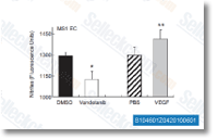 Click to enlarge | Rating | |
| Source | hypertension 2011 58, 85-92. Vandetanib (ZD6474) purchased from Selleck | |
| Method | NO Assay | |
| Cell Lines | MS1 endothelial cells | |
| Concentrations | 1 μmol/L | |
| Incubation Time | 1.5 h | |
| Results | Incubation of vandetanib with MS1 EC significantly decreased nitrite production compared with the matched vehicle (DMSO;Figure 2; P=0.0003). These findings demonstrate that vandetanib lowered EC NO levels. |
Kinase fusions are frequent in Spitz tumours and spitzoid melanomas [Wiesner T, et al. Nat Commun 2014;5:3116]
Contrary effects of the receptor tyrosine kinase inhibitor Vandetanib on constitutive and flow-stimulated nitric oxide elaboration in humans. [Mayer EL, et al. Hypertension 2011;58(1):85-92]
Ponatinib is a potent inhibitor of wild-type and drug-resistant gatekeeper mutant RET kinase. [Mologni L, et al. Mol Cell Endocrinol 2013;377(1-2):1-6]
