
Antibodies


Tubastatin A is a potent and selective HDAC6 inhibitor with IC50 of 15 nM. It is selective against all the other isozymes (1000-fold) except HDAC8 (57-fold).
| Size | Price | Stock | Quantity | ||||
|---|---|---|---|---|---|---|---|
| |||||||
 Distributor Tel: +886-2-2827-1197jimmy@new.abscience.com.tw
| Description | Tubastatin A is a potent and selective HDAC6 inhibitor with IC50 of 15 nM. It is selective against all the other isozymes (1000-fold) except HDAC8 (57-fold). | |||||
|---|---|---|---|---|---|---|
| Targets | HDAC6 | |||||
| IC50 | 15 nM [1] | |||||
| In vitro | Tubastatin A is selective at all isozymes except HDAC8 and maintains over 1000-fold selectivity against all isoforms excluding HDAC8, where it has approximately 57-fold selectivity. Tubastatin A preferentially induces α-tubulin hyperacetylation at 2.5 μM. Slight induction of histone hyperacetylation is seen for Tubastatin A at 10 μM. Tubastatin A displays dose-dependent protection against homocysteic acid-induced neuronal cell death starting at 5 μM with near complete protection at 10 μM. [1] Tubastatin A (10 μM) induces an increase in acetylated-α-tubulin levels and the restoration of primary cilia expression in the cholangiocarcinoma cell lines (18-fold); and the restoration of primary cilia correlated with downregulated Hedgehog (Hh) and MAPK signaling pathways, as well as decreased cell proliferation rates (in average by 50%) and invasion (by 40%). [2]Tubastatin A shows significant inhibition of TNF-α and IL-6 in LPS stimulated human THP-1 macrophages with an IC50 of 272 nM and 712 nM. Tubastatin A inhibits nitric oxide (NO) secretion in murine Raw 264.7 macrophages dose depenndently with an IC50 of 4.2 μM. [3] | |||||
| In vivo | Tubastatin A reduces the growth of cholangiocarcinoma in vivo. Tubastatin A (10 mg/kg) induces a 6-fold lower mean tumor weights in syngeneic rat orthotopic model of cholangiocarcinoma, and reduction of the ratios of tumor weight to liver weight and body weight (5- and 5.6-fold, respectively), as well as a greater frequency of ciliated cholangiocytes compared with controls (29% vs 1.4%). Tubastatin A significantly decreases the amount of PCNA-positive cells in the treated tumors compared with vehicle controls (34% vs 65%). [2] Tubastat A shows significant inhibition of paw volume at 30 mg/kg i.p. in a Freund’s complete adjuvant (FCA) induced animal model of inflammation. Tubastat A (30 mg/kg i.p.) significant attenuates clinical scores (~ 70%), and IL-6 expression in paw tissues of collagen induced arthritis DBA1 mouse. [3] | |||||
| Features | ||||||
| HDAC enzymatic assays | Tubastatin A is dissolved and diluted in assay buffer (50 mM HEPES, pH 7.4, 100 mM KCl, 0.001% Tween-20, 0.05% BSA, and 20 μM tris(2-carboxyethyl)phosphine) to 6-fold of the final concentration. HDAC enzymes are diluted to 1.5-fold of the final concentration in assay buffer and pre-incubated with Tubastatin A for 10 minutes before the addition of the substrate. The amount of FTS (HDAC1, HDAC2, HDAC3, and HDAC6) or MAZ-1675 (HDAC4, HDAC5, HDAC7, HDAC8, and HDAC9) used for each enzyme is equal to the Michaelis constant (Km), as determined by a titration curve. FTS or MAZ-1675 is diluted in assay buffer to 6-fold the final concentration with 0.3 μM sequencing grade trypsin. The substrate/trypsin mix is added to the enzyme/compound mix and the plate is shaken for 60 seconds and then placed into a SpectraMax M5 microtiter plate reader. The enzymatic reaction is monitored for release of 7-amino-4-methoxy-coumarin over 30 minutes, after deacetylation of the lysine side chain in the peptide substrate, and the linear rate of the reaction is calculated. |
|---|
| Cell lines | Human cholangiocarcinoma cell lines HuCCT-1 |
|---|---|
| Concentrations | ~10 μM |
| Incubation Time | 21 days |
| Method | Anchorage-independent growth is assessed by growing cells in soft agar. About 25,000 cells suspended in 0.4% agar in culture media are layered over a 1% agar layer in a 6-well plate. Media are added twice a week and pictures are taken after 21 days of incubation. The number and size of colonies are analyzed using the Gel-Pro software. |
| Animal Models | Rat cholangiocarcinoma xenografts BDEneu |
|---|---|
| Dosages | 10 mg/kg |
| Administration | i.p. daily |
| Molecular Weight (MW) | 335.4 |
|---|---|
| Formula | C20H21N3O2 |
| CAS No. | 1252003-15-8 |
| Storage | 3 years -20℃Powder |
|---|---|
| 6 months-80℃in DMSO | |
| Syonnyms | N/A |
| Solubility (25°C) * | In vitro | DMSO | 9 mg/mL (26 mM) |
|---|---|---|---|
| Water | <1 mg/mL (<1 mM) | ||
| Ethanol | <1 mg/mL (<1 mM) | ||
| * <1 mg/ml means slightly soluble or insoluble. * Please note that Selleck tests the solubility of all compounds in-house, and the actual solubility may differ slightly from published values. This is normal and is due to slight batch-to-batch variations. | |||
| Chemical Name | N-hydroxy-4-((2-methyl-2,3,4,5-tetrahydro-1H-indeno[1,2-c]pyridin-5-yl)methyl)benzamide |
|---|
 Click to enlarge | Rating | |
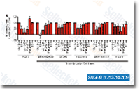
| Source | J Biol Chem. 2013 May 17;288(20):14400-7 . Tubastatin A purchased from Selleck | |
| Method | qRT-PCR | |
| Cell Lines | Triple-Negative Cancer Lines | |
| Concentrations | 1 uM | |
| Incubation Time | 24 h | |
| Results | Three of the five cell lines (MDA- MB-468, BT549, and HCC1937) showed suppression of mesenchymal gene expression following treatment with TSA, MS-275, or LBH589, and the other two (MDA-MB-231 and Hs578T) had a more modest reduction in mesenchymal gene expression. *p<0.05. Student’s t test for mean of mesenchymal gene markers, for each drug-treated cell line compared with control DMSO-treated cell line. |
Histone Deacetylase Inhibitors (HDACis) That Release the Positive Transcription Elongation Factor b (P-TEFb) from Its Inhibitory Complex Also Activate HIV Transcription. [Bartholomeeusen K, et al. J Biol Chem 2013;288(20):14400-7]
Dynamic Chromatin Modification Sustains Epithelial-Mesenchymal Transition following Inducible Expression of Snail-1. [Javaid S ,et al. Cell Rep 2013;5(6):1679-89]
