
Antibodies


S3I-201 shows potent inhibition of STAT3 DNA-binding activity with IC50 of 86 μM, and low activity towards STAT1 and STAT5.
| Size | Price | Stock | Quantity | ||||||||||||||||
|---|---|---|---|---|---|---|---|---|---|---|---|---|---|---|---|---|---|---|---|
| |||||||||||||||||||
 Distributor Tel: +886-2-2827-1197jimmy@new.abscience.com.tw
| Description | S3I-201 shows potent inhibition of STAT3 DNA-binding activity with IC50 of 86 μM, and low activity towards STAT1 and STAT5. | |||||
|---|---|---|---|---|---|---|
| Targets | Stat3 | |||||
| IC50 | 86 μM [1] | |||||
| In vitro | S3I-201 inhibits growth and induces apoptosis preferentially in tumor cells that contain persistently activated Stat3 by inhibiting Stat3·Stat3 complex formation and Stat3 DNA-binding and transcriptional activitie. Moreover, S3I-201 also inhibits the expression of the Stat3-regulated genes encoding cyclin D1, Bcl-xL, and survivin. [1]S3I-201 inhibits breast carcinoma MDA-MB-435, MDA-MB-453 and MDA-MB-231 cell lines with IC50 of 100 μM. In addition, the cells with impaired TGF-β signaling are four times as sensitive to the STAT3 inhibitor S3I-201.[2] A recent study shows that S3I-201 potentiates the antiproliferative effect of cetuximab in HepG2 and Huh-7 cells via the STAT3 signalling pathway. [3] | |||||
| In vivo | S3I-201 (5 mg/kg, i.v. every 2 or every 3 days) shows the antitumor efficacy in mouse models with human breast tumor xenografts that harbor constitutively active Stat3. [1] S3I-201 treatment reduces Varicella-zoster virus (VZV) replication on the basis of the bioluminescence signal and the number of positive skin xenografts compared with DMSO-treated mice by inhibiting STAT3 phosphorylation. [4] | |||||
| Features | A chemical probe inhibitor of Stat3 activity. | |||||
| In vitro Stat3 DNA-binding assay and EMSA analysis | Briefly, 100 mL of biotinyl-e-Ac-EPQpYEEIEL-OH (in 50 mM Tris/150 mM NaCl, pH 7.5) is added to each well of streptavidin-coated 96-well microtiter plates and incubated with shaking at 4 °C overnight. Then plates are rinsed with PBS/Tween 20 and then two times with 200 mL of BSA-T-PBS (0.2% BSA/0.1% Tween 20/PBS). Then 50 mL of Lck-SH2-GST fusion protein (6.4 ng/ml in BSA-T-PBS) is added to each well of the 96-well plate in the presence and absence of 50 mL of S3I-201 (for 30 and 100 mM final concentrations), and the plate is shaken at room temperature for 4 hours. After solutions are removed, each well is rinsed four times with BSA-T-PBS (200 mL), and 100 mL of polyclonal rabbit anti-GST antibody (100 ng/mL in BSA-T-PBS) is added to each well and incubated at 4 °C overnight. After washing with BSA-T-PBS, 100 mL of 200 ng/mL BSA-T-PBS horseradish peroxidase-conjugated mouse anti-rabbit antibody is added to each well and incubated for 45 minutes at room temperature. After four washing steps with BSA-T-PBS and three washing steps with PBS-T, 100 mL of peroxidase substrate is added to each well and incubated for 5-15 minutes. The peroxidase reaction is stopped by adding 100 mL of 1 M sulfuric acid solution, and absorbance is read at 450 nm with an ELISA plate rea |
|---|
| Cell lines | MDA-MB-435, MDA-MB-453 and MDA-MB-231 cells lines |
|---|---|
| Concentrations | ~ 250 μM |
| Incubation Time | 72 hours |
| Method | The MTT assay is based on the conversion of the yellow tetrazolium salt MTT to purple formazan crystals by metabolically active cells. The MTT assay provides a quantitative determination of viable cells. Cells are seeded in 96-well microplates in complete culture medium in the absence or presence of increasing serial dosages of S3I-201 as indicated. At 72 hours after culture, the number of viable cells is measured by adding 100 μL/well of 2 mg/mL MTT solution. After 2 hours, the medium is removed, and the formazan crystals are dissolved by adding 100 μL dimethylsulfoxide per well. The absorbance is read at 590 nm with an enzyme-linked immunosorbent assay reader. Each treatment point is performed in 10 wells or sextuplicate. |
| Animal Models | Human breast cancer MDA-MB-231 cells are injected s.c. into the left flank of athymic nu/nu mice. |
|---|---|
| Dosages | ≤5 mg/kg |
| Administration | Administered via i.v. |
| Solubility | 1% DMSO/30% polyethylene glycol/1% Tween 80, 15 mg/mL |
| Molecular Weight (MW) | 365.36 |
|---|---|
| Formula | C16H15NO7S |
| CAS No. | 501919-59-1 |
| Storage | 3 years -20℃Powder |
|---|---|
| 6 months-80℃in DMSO | |
| Syonnyms | N/A |
| Solubility (25°C) * | In vitro | DMSO | 73 mg/mL (199 mM) |
|---|---|---|---|
| Water | <1 mg/mL (<1 mM) | ||
| Ethanol | <1 mg/mL (<1 mM) | ||
| In vivo | 1% DMSO/30% polyethylene glycol/1% Tween 80, | 15 mg/mL | |
| * <1 mg/ml means slightly soluble or insoluble. * Please note that Selleck tests the solubility of all compounds in-house, and the actual solubility may differ slightly from published values. This is normal and is due to slight batch-to-batch variations. | |||
| Chemical Name | 2-hydroxy-4-(2-(tosyloxy)acetamido)benzoic acid |
|---|
| Stock Solution (1ml DMSO) | 1mM | 10mM | 20mM | 30mM |
|---|---|---|---|---|
| Mass(mg) | 0.36536 | 3.6536 | 7.3072 | 10.9608 |
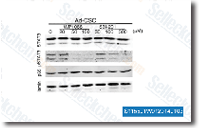 Click to enlarge | Rating | |
| Source | J Biol Chem 2013 288(36):26167-76. S3I-201 purchased from Selleck | |
| Method | Western blot | |
| Cell Lines | adherent glioma CSCs | |
| Concentrations | 30, 100, 300 uM | |
| Incubation Time | 2 h | |
| Results | Both WP1066 and S3I-201 were extremely effective in inhibiting STAT3 tyrosine phosphorylation in CSCs, with nearly complete inhibition of pSTAT3 at 50 μM WP1066 and 300 μM S3I-201. |
Inhibiting EGF receptor or SRC family kinase signaling overcomes BRAF inhibitor resistance in melanoma. [Girotti MR, et al. Cancer Discov 2012;3(2):158-67]
Suppression of signal transducers and activators of transcription 1 in hepatocellular carcinoma is associated with tumor progression. [Hosui A, et al. Int J Cancer 2012;131(12):2774-84]
Critical role of the WASF3 gene in JAK2/STAT3 regulation of cancer cell motility. [Teng Y, et al. Carcinogenesis 2013;34(9):1994-9]
