
Antibodies


RAF265 (CHIR-265) is a potent selective inhibitor of C-Raf/B-Raf/B-Raf V600E with IC50 of 3-60 nM, and exhibits potent inhibition onVEGFR2 phosphorylation with EC50 of 30 nM. Phase 2.
| Size | Price | Stock | Quantity | ||||||||||||||||||||
|---|---|---|---|---|---|---|---|---|---|---|---|---|---|---|---|---|---|---|---|---|---|---|---|
| |||||||||||||||||||||||
 Distributor Tel: +886-2-2827-1197jimmy@new.abscience.com.tw
| Description | RAF265 (CHIR-265) is a potent selective inhibitor of C-Raf/B-Raf/B-Raf V600E with IC50 of 3-60 nM, and exhibits potent inhibition on VEGFR2 phosphorylation with EC50 of 30 nM. Phase 2. | |||||
|---|---|---|---|---|---|---|
| Targets | B-Raf | VEGFR2 | ||||
| IC50 | 3-60 nM | 30 nM (EC50) [1] | ||||
| In vitro | RAF265 inhibits C-Raf, wild type B-Raf and mutant (V600E) B-Raf. RAF265 effectively block phosphorylation of Rafs downstream substrates MEK and ERK in cells and also kill melanoma and colorectal cancer cell lines harboring B-Raf mutations independent of PTEN mutation status. Raf kinase inhibition by RAF265 in mutant B-Raf melanoma cell lines causes cell cycle arrest and induces apoptosis, mimicking the effect of Raf RNAi in these cells. RAF265 also potently inhibits the phosphorylation of VEGFR2 and proliferation of VEGF-stimulated hMVEC. [1] In HT29 and MDAMB231 cells, RAF265 shows inhibitory activity with IC20 of 1 to 3 μM and IC50 of 5 to 10 μM, respectively. While RAF265 leads to a significant decrease in clonogenic survival in all tested cell lines, which means that RAF265 induces a dominant effect on clonogenic survival. Addition of RAF265 to RAD001 in HCT116 cells could lead to moderately decreased AKT, S6 protein, and 4EBP1 phosphorylation. [2]Raf265 markedly reduces the protein level of Bcl-2 and great inhibitory in CM- and NCI-H727 cells, while having no effect on the TRAIL susceptibility of BON1 and GOT1 cells. [3] Protein kinase D3 (PRKD3) that when knocked down could enhance cell killing by RAF265 in A2058 melanoma cells, which prevent reactivation of MAPK signaling, induce PARP cleavage, increase caspase activity, interrupt cell-cycle progression, and inhibit colony formation. [4] | |||||
| In vivo | RAF265 shows 71% to 72% TVI% (tumor volume inhibition percentage) in HCT116 xenografts at 12 mg/kg. While the combination of RAF265 and RAD001 shows enhanced antitumor activity with increased T10 (time to achieve a relative tumor volume of 10 times the initial tumor volume) and tumor growth delay. The combination of RAD001 and RAF265 also significantly enhances the activation of caspase-3 in HCT116 and MDAMB231 but not in A549 xenografts. [2] RAF265 inhibits FDG (2-deoxy-2-[18F]fluoro-d-glucose) accumulation and decreases the tumor volumes in A375M xenografts by orally dosed of 100 mg/kg. [5] | |||||
| Features | ||||||
| Assay Protocol | Raf and Mek are combined at 2 × final concentrations in assay buffer (50 mM Tris, pH 7.5, 15 mM MgCl2. 0.1 mM EDTA and 1 mM DTT) and dispensed 15 μL per well in polypropylene assay plates. Background levels are determined in wells containing Mek and DMSO without Raf. To the Raf/Mek containing wells is added 3 μL of 10 × of RAF265 diluted in 100% DMSO. The raf kinase activity reaction is started by the addition of 12 μL per well of 2.5 × 33P-ATP diluted in assay buffer. After 45-60 minutes, the reactions are stopped with the addition of 70 μL of stop reagent (30 mM EDTA). Filtration plates are pre-wetted for 5 min with 70% ethanol, and then rinsed by filtration with wash buffer. Samples (90 μL) from the reaction wells are then transferred to the filtration plates. The filtration plates are washed 6 × with wash buffer using Millipore filtration apparatus. The plates are dried and 100 μL per well of scintillation fluid is added. The CPM is then determined using a Wallac Microbeta 1450 reader. |
|---|---|
| In Vitro Raf Screen | The activity of various isoforms of Raf serine/threonine kinases can be measured by providing ATP, MEK substrate, and assaying the transfer of phosphate moiety to the MEK residue. Recombinant isoforms of Raf are obtained by purification from sf9 insect cells infected with a human Raf recombinant baculovirus expression vector. Recombinant kinase inactive MEK is expressed in E. coli and labeled with Biotin post purification. For each assay, RAF265 is serially diluted in DMSO then mixed with Raf (0.50 nM) and kinase inactive biotin-MEK (50 nM) in reaction buffer plus ATP (1 uM). Reactions are subsequently incubated for 2 hours at room temperature and stopped by the addition of 0.5 M EDTA. Stopped reaction mixture is transferred to a neutradavin-coated plate and incubated for 1 hour. Phosphorylated product is measured with the DELFIA time-resolved fluorescence system, using a rabbit anti-p-MEK as the primary antibody and europium labeled anti-rabbit as the secondary antibody. Time resolved fluorescence is read on a Wallac 1232 DELFIA fluorometer. The concentration of RAF265 for 50% inhibition is calculated by non-linear regression using XL Fit data analysis software. |
| Cell lines | Human A549 and H460 lung, HT29 and HCT 116 colon, and MDAMB231 breast cancer cell lines |
|---|---|
| Concentrations | 0.1 - 10 μM |
| Incubation Time | 48 hours |
| Method | The MTT assay and Bliss additivism model are used to assess the effect of RAF265 on cell viability. In each well of a 96-well plate, 1 × 10⁴ cells are grown in 200 μL of medium. After 24 hours, RAF265 is added to achieve a final concentration of 0.1 to 10 μM. After 48 hours of treatment, 20 μL of 5 mg/mL MTT solution in PBS is added to each well. After 4 hours, supernatant is removed and formazan crystals are discarded in 200 μL of DMSO. Absorbance is then measured at 595 nm using an absorbance plate reader. Data are expressed as the percentage of viable cells. |
| Animal Models | A549, H460, HCT116, or MDAMB231 cells are injected s.c. into the flank region of 6-wk-old female athymic mice. |
|---|---|
| Dosages | 12 mg/kg |
| Administration | Orally administered daily |
| Solubility | 30% PEG400/0.5% Tween80/5% propylene glycol, 30 mg/mL |
| NCT Number | Recruitment | Conditions | Sponsor /Collaborators | Start Date | Phases |
|---|---|---|---|---|---|
| NCT00304525 | Active, not recruiting | Metastatic Melanoma | Novartis|Novartis Pharmaceuticals | 2006-04 | Phase 2 |
| NCT01352273 | Active, not recruiting | Advanced Solid Tumors | Novartis|Novartis Pharmaceuticals | 2011-06 | Phase 1 |
Download RAF265 (CHIR-265) SDF
| Molecular Weight (MW) | 518.41 |
|---|---|
| Formula | C24H16F6N6O |
| CAS No. | 927880-90-8 |
| Storage | 3 years -20℃Powder |
|---|---|
| 6 months-80℃in DMSO | |
| Syonnyms | N/A |
| Solubility (25°C) * | In vitro | DMSO | 100 mg/mL (192 mM) |
|---|---|---|---|
| Water | <1 mg/mL (<1 mM) | ||
| Ethanol | 33 mg/mL (63 mM) | ||
| In vivo | 30% PEG400/0.5% Tween80/5% propylene glycol, | 30 mg/mL | |
| * <1 mg/ml means slightly soluble or insoluble. * Please note that Selleck tests the solubility of all compounds in-house, and the actual solubility may differ slightly from published values. This is normal and is due to slight batch-to-batch variations. | |||
| Chemical Name | 1-methyl-5-(2-(5-(trifluoromethyl)-1H-imidazol-2-yl)pyridin-4-yloxy)-N-(4-(trifluoromethyl)phenyl)-1H-benzo[d]imidazol-2-amine |
|---|
| Stock Solution (1ml DMSO) | 1mM | 10mM | 20mM | 30mM |
|---|---|---|---|---|
| Mass(mg) | 0.51841 | 5.1841 | 10.3682 | 15.5523 |
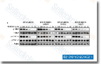 Click to enlarge | Rating | |
| Source | Pigment Cell Melanoma Res, 2014, 27(1):124-33. RAF265 (CHIR-265) purchased from Selleck | |
| Method | Western blot | |
| Cell Lines | A375 cells | |
| Concentrations | 0-60 uM | |
| Incubation Time | 24 h | |
| Results | Immunoblots showing levels of phospho-MEK (p-MEK), total MEK (t-MEK), phospho-ERK1/2 (p-ERK1/2) and total ERK1/2 (t-ERK1/2) in A375 cells transduced with a retrovirus expressing BRAFV600E, BRAFV600E/L505H, BRAFV600E/F516G or BRAFV600E/T529N and treated with increasing doses of RAF265. α-tubulin (TUBA) was monitored as a loading control. |
Cyclic AMP/PKA-dependent Paradoxical Activation of Raf/MEK/ERK Signaling in Polycystin-2 Defective Mice Treated with Sorafenib. [Carlo Spirli, et al. Hepatology 2012;56(6):2363-74]
Resistance to vemurafenib resulting from a novel mutation in the BRAFV600E kinase domain. [Wagenaar TR, et al. Pigment Cell Melanoma Res 2013;27(1):124-33]
