
Antibodies


Paclitaxel is a microtubule polymer stabilizer with IC50 of 0.1 pM in human endothelial cells.
| Size | Price | Stock | Quantity | ||||||||||||
|---|---|---|---|---|---|---|---|---|---|---|---|---|---|---|---|
| |||||||||||||||
 Distributor Tel: +886-2-2827-1197jimmy@new.abscience.com.tw
| Description | Paclitaxel is a microtubule polymer stabilizer with IC50 of 0.1 pM in human endothelial cells. | |||||
|---|---|---|---|---|---|---|
| Targets | Microtubule (human endothelial cells) | |||||
| IC50 | 0.1 pM [1] | |||||
| In vitro | Paclitaxel inhibits non-endothelial type human cells at 104 - to 105 -fold higher concentrations, with IC50 of 1 nM-10 nM. The selectivity of Paclitaxel inhibition of cell proliferation is also species specific, as mouse ECs are not sensitive to Paclitaxel at ultra low concentrations. Inhibition of human ECs by Paclitaxel at ultra low concentrations does not affect the cellular microtubule structure, and the treated cells do not show G2/M cell cycle arrest and apoptosis, suggesting a novel but as yet unidentified mechanism of action. In an in vitro angiogenesis assay, Paclitaxel at ultra low concentrations blocks human ECs from forming sprouts and tubes in the three-dimensional fibrin matrix. [1] In the presence of SMF, the efficient concentration of Paclitaxel on K562 cells is decreased from 50 to 10 ng/mL. The cell cycle arrest effect of Paclitaxel with or without SMF on K562 cells is correlated with DNA damage. [2] Paclitaxel alone causes a time-dependent inhibition of CDK1 in four cell lines including A549 cells, H358, H1395 cells and H1666 cells. [3] | |||||
| In vivo | The inhibition rations of Paclitaxel alone on BC-V and BC-ER tumors are 49.78% and 51.23%, respectively. Treatment of six cycles of 20 mg/kg Paclitaxel significantly reduces the percentages of Ki-67-positive cells to 20.4% in BC-V tumors and 25.1% in BC-ER tumors, respectively. [4] | |||||
| Features | ||||||
| Cell lines | Cells including human neonatal dermal microvascular ECs (HMVECs), human umbilical vein ECs (HUVECs), human umbilical artery ECs (HUAVECs), normal human astrocytes (NHAs), normal human dermal fibroblasts (NHDFs), normal human epidermal keratinocytes (NHEKs), human mammary epithelial cells (HMEpCs), human prostate epithelial cells (PrEpCs) and human umbilical artery smooth muscle cells (UASMCs) |
|---|---|
| Concentrations | 0.1-100 pM |
| Incubation Time | 72 hours |
| Method | Cells including human neonatal dermal microvascular ECs (HMVECs), human umbilical vein ECs (HUVECs), human umbilical artery ECs (HUAVECs), normal human astrocytes (NHAs), normal human dermal fibroblasts (NHDFs), normal human epidermal keratinocytes (NHEKs), human mammary epithelial cells (HMEpCs), human prostate epithelial cells (PrEpCs) and human umbilical artery smooth muscle cells (UASMCs) are cultured. Cell proliferations are performed in 96-well plates using cells between passages 6 and 12. Cells are seeded at 3000–5000 cells/well and allowed to attach for 4 hours. Paclitaxel, diluted in culture medium, is added in quadruplicate wells and the cells ae incubated for 3 days before MTS reagents are added to quantitate the live cells in each well. |
| Animal Models | Female, 20-22 g homozygous nude athymic mice with BC-V and BC-ER tumors |
|---|---|
| Dosages | 20 mg/kg |
| Administration | Administered via i.v. |
| Solubility | 1% DMSO/30% polyethylene glycol/1% Tween 80, 30 mg/mL |
| NCT Number | Recruitment | Conditions | Sponsor /Collaborators | Start Date | Phases |
|---|---|---|---|---|---|
| NCT01993810 | Not yet recruiting | Stage IIA Non-small Cell Lung Cancer|Stage IIB Non-small Cell Lung Cancer|Stage IIIA Non-small Cell Lung Cancer|Stage IIIB Non-small Cell Lung Cancer | Radiation Therapy Oncology Group|National Cancer Institute (NCI) | 2014-02 | Phase 3 |
| NCT02018458 | Not yet recruiting | Breast Cancer | Baylor Research Institute | 2014-03 | Phase 1|Phase 2 |
| NCT01969578 | Not yet recruiting | Salivary Gland Cancer | European Organisation for Research and Treatment of Cancer - EORTC | 2014-03 | Phase 2 |
| NCT02012192 | Not yet recruiting | Epithelial Ovarian Cancer|Fallopian Tube Cancer|Primary Peritoneal Cancer | Medical University Innsbruck|European Commission | 2014-06 | Phase 1|Phase 2 |
| NCT01867086 | Not yet recruiting | Stage III Ovarian Cancer|Stage IV Ovarian Cancer | Gradalis, Inc. | 2014-06 | Phase 2 |
| Molecular Weight (MW) | 853.91 |
|---|---|
| Formula | C47H51NO14 |
| CAS No. | 33069-62-4 |
| Storage | 3 years -20℃Powder |
|---|---|
| 6 months-80℃in DMSO | |
| Syonnyms | N/A |
| Solubility (25°C) * | In vitro | DMSO | 171 mg/mL (200 mM) |
|---|---|---|---|
| Water | <1 mg/mL (<1 mM) | ||
| Ethanol | 18 mg/mL (21 mM) | ||
| In vivo | 1% DMSO/30% polyethylene glycol/1% Tween 80, | 30 mg/mL | |
| * <1 mg/ml means slightly soluble or insoluble. * Please note that Selleck tests the solubility of all compounds in-house, and the actual solubility may differ slightly from published values. This is normal and is due to slight batch-to-batch variations. | |||
| Chemical Name | Benzenepropanoic acid, β-(benzoylamino)-α-hydroxy-, (2aR,4S,4aS,6R,9S,11S,12S,12aR,12bS)-6,12b-bis(acetyloxy)-12-(benzoyloxy)-2a,3,4,4a,5,6,9,10,11,12,12a,12b-dodecahydro-4,11-dihydroxy-4a,8,13,13-tetramethyl-5-oxo-7,11-methano-1H-cyclodeca[3,4]benz[1,2-b]oxet-9-yl ester, (αR,βS)- |
|---|
| Stock Solution (1ml DMSO) | 1mM | 10mM | 20mM | 30mM |
|---|---|---|---|---|
| Mass(mg) | 0.85391 | 8.5391 | 17.0782 | 25.6173 |
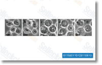 Click to enlarge | Rating | |
| Source | RSC Adv 2011 1, 884-892. Paclitaxel purchased from Selleck | |
| Method | Microscope image | |
| Cell Lines | U937 cells | |
| Concentrations | ||
| Incubation Time | 24 h | |
| Results | The ODS-treated cells in Fig. b looked identical to the untreated cells in Fig. a with a similar rate of natural cell death of less than 3%. When Taxol microcrystals are added to the cell culture (the standard way that Taxol is released into biological systems), decreased cell proliferation rate, pronounced swelling and vacuolization are observed, as shown in Fig. c. |
TGFβ/TNFα-Mediated Epithelial-Mesenchymal Transition Generates Breast Cancer Stem Cells with a Claudin-Low Phenotype. [Suwanabol PA, et al. Am J Physiol Heart Circ Physiol 2012;71(13):4707-19]
The downregulation of Mcl-1 via USP9X inhibition sensitizes solid tumors to Bcl-xl inhibition. [Peddaboina C, et al. BMC Cancer 2012;12:541]
Development, validation and pilot screening of an in vitro multi-cellular three-dimensional cancer spheroid assay for anti-cancer drug testing. [Rati Lama, et al. Bioorg Med Chem 2012;21(4):922-31]
