
Antibodies


MK-2206 2HCl is a highly selective inhibitor of Akt1/2/3 with IC50 of 8 nM/12 nM/65 nM, respectively; no inhibitory activities against 250 other protein kinases observed. Phase 2.
| Size | Price | Stock | Quantity | ||||||||||||||||||||
|---|---|---|---|---|---|---|---|---|---|---|---|---|---|---|---|---|---|---|---|---|---|---|---|
| |||||||||||||||||||||||
 Distributor Tel: +886-2-2827-1197jimmy@new.abscience.com.tw
| Description | MK-2206 2HCl is a highly selective inhibitor of Akt1/2/3 with IC50 of 8 nM/12 nM/65 nM, respectively; no inhibitory activities against 250 other protein kinases observed. Phase 2. | |||||
|---|---|---|---|---|---|---|
| Targets | Akt1 | Akt2 | Akt3 | |||
| IC50 | 8 nM | 12 nM | 65 nM [1] | |||
| In vitro | MK-2206 is an allosteric inhibitor and is activated by the pleckstrin homology domain. MK-2206 inhibits auto-phosphorylation of both Akt T308 and S473. MK-2206 also prevents Akt-mediated phosphorylation of downstream signaling molecules, including TSC2, PRAS40 and ribosomal S6 proteins. [1] MK-2206 inhibits Ras wild-type (WT) cell lines (A431, HCC827, and NCI-H292) more potently when compared to Ras-mutant cell lines (NCI-H358, NCI-H23, NCI-H1299, and Calu-6). MK-2206 also shows synergistic responses in combination with cytotoxic agents such as erlotinib or lapatinib in lung NCI-H460 or ovarian A2780 tumor cells.[2] MK-2206 or siRNA-mediated Akt inhibition strongly activates autophagy in human glioma cells. However, eukaryotic elongation factor-2 (eEF-2) silencing suppresses MK-2206-induced-autophagy, with a promotion of apoptotic cell death. [3] | |||||
| In vivo | MK-2206 shows 60% TGI and inhibits more than 70 % of phospho-Akt1/2 (T308 and S473) in A2780 ovarian cancer xenografts at a dose of 240 mg/kg. [1] MK-2206 exhibits significant antitumor activity in NCI-H292 xenograft in combination with erlotinib or lapatinib. [2] | |||||
| Features | The first allosteric small molecule inhibitor of Akt to enter clinical development. | |||||
| Akt kinases assay | Akt kinases are assayed by a GSK-derived biotinylated peptide substrate. The extent of peptide phosphorylation is determined by Homogeneous Time Resolved Fluorescence (HTRF) using a lanthanide chelate (Lance)-coupled monoclonal antibody specific for the phosphopeptide in combination with a streptavidin-linked allophycocyanin (SA-APC) fluorophore which will bind to the biotin moiety on the peptide. When the Lance and APC are in proximity, a non-radiative energy transfer takes place from the Lance to the APC, followed by emission of light from APC at 655 nm. Working Solution: 100X protease inhibitor cocktail (PIC): 1mg/mL benzamidine, 0.5 mg/mL pepstatin, 0.5 mg/mL leupeptin, 0.5 mg/mL aprotinin; 10X assay buffer: 500 mM HEPES, pH7.5, 1% PEG, 16.6 mM EDTA, 1 mM EGTA, 1% BSA, 20 mM 9-glycerol phosphate; Quench buffer 50 mM HEPES pH 7.3, 16.6 mM EDTA, 0.1% BSA, 0.1% Triton X-100, 0.17 nM labeled monoclonal antibody, 0.0067 mg/mL SA-APC; ATP/MgCl2 working solution: 1X Assay buffer, 1 mM DTT, 1X PIC, 5% glycerol, active Akt; Peptide working solution: 1X Assay buffer, 1 mM DTT, 1X PIC, 5% glycerol, 2 TM GSK biotinylated peptide. The reaction is assembled by adding 16 µL of ATP/MgCl2 working solution to the appropriate wells. MK-2206 or vehicle (1.0 µL) is added followed by 10 µL of peptide working solution. The reaction is started by adding 13 μL of the enzyme working solution and mixing. The reaction is allowed to proceed for 50 min and then stopped by the addition of 60 µL HTRF quench buffer. The stopped reactions are incubated at room temperature for at least 30 min and then read in the instrument. |
|---|
| Cell lines | A431, HCC827, NCI-H292, NCI-H358, NCI-H23, NCI-H1299, Calu-6 and NCI-H460 cells |
|---|---|
| Concentrations | 0, 0.3, 1 and 3 μM |
| Incubation Time | 72 or 96 hours |
| Method | MK-2206 is dissolved in DMSO as a stock solution and diluted by culture media before use. Cells are seeded at a density of 2-3 × 10³ in 96-well plates and incubated for 24 hours. Then MK-2206 (0, 0.3, 1 and 3 μM) is added to the cells. Cell proliferation is determined after 72 or 96 hours. |
| Animal Models | SK-OV-3, NCI-H292, HCC70, PC-3, and NCI-H460 models in male CD1-nude mice |
|---|---|
| Dosages | 120 mg/kg |
| Administration | Orally administered |
| Solubility | 15% Captisol, 17 mg/mL |
| NCT Number | Recruitment | Conditions | Sponsor /Collaborators | Start Date | Phases |
|---|---|---|---|---|---|
| NCT01783171 | Recruiting | Acinar Cell Adenocarcinoma of the Pancreas|Duct Cell Adenocarcinoma of the Pancreas|Recurrent Pancreatic Cancer|Stage III Pancreatic Cancer|Stage IV Pancreatic Cancer | National Cancer Institute (NCI) | 2013-01 | Phase 1 |
| NCT01776008 | Recruiting | Estrogen Receptor-positive Breast Cancer|HER2-negative Breast Cancer|Recurrent Breast Cancer|Stage II Breast Cancer|Stage IIIA Breast Cancer|Stage IIIB Breast Cancer|Stage IIIC Breast Cancer | National Cancer Institute (NCI) | 2013-01 | Phase 2 |
| NCT01859182 | Terminated | Adenocarcinoma of the Gallbladder|Adenocarcinoma With Squamous Metaplasia of the Gallbladder|Adult Primary Cholangiocellular Carcinoma|Advanced Adult Primary Liver Cancer|Cholangiocarcinoma of the Ext | National Cancer Institute (NCI) | 2013-01 | Phase 2 |
| NCT01802320 | Recruiting | Mucinous Adenocarcinoma of the Colon|Mucinous Adenocarcinoma of the Rectum|Recurrent Colon Cancer|Recurrent Rectal Cancer|Signet Ring Adenocarcinoma of the Colon|Signet Ring Adenocarcinoma of the Rect | National Cancer Institute (NCI) | 2013-03 | Phase 2 |
| NCT01714128 | Not yet recruiting | Breast Cancer | Washington University School of Medicine | 2013-06 |
| Molecular Weight (MW) | 480.39 |
|---|---|
| Formula | C25H21N5O.2HCl |
| CAS No. | 1032350-13-2 |
| Storage | 3 years -20℃Powder |
|---|---|
| 6 months-80℃in DMSO | |
| Syonnyms | N/A |
| Solubility (25°C) * | In vitro | DMSO | 14 mg/mL (29 mM) |
|---|---|---|---|
| Water | 1 mg/mL (2 mM) | ||
| Ethanol | <1 mg/mL (<1 mM) | ||
| In vivo | 15% Captisol, | 17 mg/mL | |
| * <1 mg/ml means slightly soluble or insoluble. * Please note that Selleck tests the solubility of all compounds in-house, and the actual solubility may differ slightly from published values. This is normal and is due to slight batch-to-batch variations. | |||
| Chemical Name | 8-(4-(1-aminocyclobutyl)phenyl)-9-phenyl-[1,2,4]triazolo[3,4-f][1,6]naphthyridin-3(2H)-one |
|---|
| Stock Solution (1ml DMSO) | 1mM | 10mM | 20mM | 30mM |
|---|---|---|---|---|
| Mass(mg) | 0.48039 | 4.8039 | 9.6078 | 14.4117 |
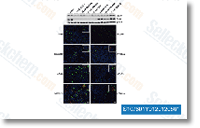 Click to enlarge | Rating | |
| Source | Biochim Biophys Acta, 2012, 1823(5), 987-96. MK-2206 2HCl purchased from Selleck | |
| Method | Immunofluorescence | |
| Cell Lines | HC11 cells | |
| Concentrations | 10 µM | |
| Incubation Time | 1 h | |
| Results | whole ADRP levels, estimated by Western blot , decreased only in the presence of MK -2206 and LY294002. In most cases, ADRP decorated the surface of small lipid droplets and appeared as little patches on large cytoplasmic lipid droplets. With the exception of SP600125, which induced a strong ADRP coating of almost all cytoplasmic lipid droplets (although this inhibitor did not increase ADRP synthesis), variation in the distribution of ADRP at the surface of lipid droplet was difficult to estimate, notably because the signal was faint and uneven. |
Direct Reversal of Glucocorticoid Resistance by AKT Inhibition in Acute Lymphoblastic Leukemia. [Piovan E, et al. Cancer Cell 2013;24(6):766-76]
Expansion of oligodendrocyte progenitor cells following SIRT1 inactivation in the adult brain. [Rafalski VA, et al. Nat Cell Biol 2013;15(6):614-24]
The Akt-SRPK-SR Axis Constitutes a Major Pathway in Transducing EGF Signaling to Regulate Alternative Splicing in the Nucleus. [Zhou Z, et al. Mol Cell 2012;47(3):422-33]