
Antibodies


MG-132 is an inhibitor of proteasome with IC50 of 100 nM, and also inhibits calpain with IC50 of 1.2 μM.
| Size | Price | Stock | Quantity | ||||||||||||||||
|---|---|---|---|---|---|---|---|---|---|---|---|---|---|---|---|---|---|---|---|
| |||||||||||||||||||
 Distributor Tel: +886-2-2827-1197jimmy@new.abscience.com.tw
| Description | MG-132 is an inhibitor of proteasome with IC50 of 100 nM, and also inhibits calpain with IC50 of 1.2 μM. | |||||
|---|---|---|---|---|---|---|
| Targets | Proteasome | |||||
| IC50 | 100 nM [1] | |||||
| In vitro | MG-132 displays >1000 times more activity than ZLLal in inhibiting the ZLLL-MCA-degrading activity of 20S proteasome with IC50 of 100 nM versus 110 μM. MG-132 also inhibits calpain with IC50 of 1.2 μM. MG-132 induces neurite outgrowth in PC12 cells at an optimal concentration of 20 nM, displaying 500 times more potency than ZLLal. [1] MG-132 (10 μM) potently inhibits TNF-α-induced NF-κB activation, interleukin-8 (IL-8) gene transcription, and IL-8 protein release in A549 cells by inhibition of proteasome-mediated IκBα degradation. [2] MG-132 treatment potently induces p53-dependent apoptosis in KIM-2 cells by 26S proteasome inhibition. [3] Unlike BzLLLCOCHO or PS-341, MG-132 treatment results in weak inhibition of the chymotrypsinlike (CT-L) and peptidylglutamyl peptide hydrolysing (PGPH) activities of the 26S proteasome, whereas multiple myeloma cells (U266 and OPM-2) are more sensitive to induction of apoptosis by MG-132 than BzLLLCOCHO. [4] MG-132 (1 μM) sensitizes TRAIL-resistant prostate cancer cells by activating the AP-1 family members c-Fos and c-Jun, which, in turn, repress the antiapoptotic molecule c-FLIP(L). [5] MG-132 significantly enhances the ability of inositol hexakisphosphate (IP6) to reduce cellular metabolic activity in both PC3 and DU145 androgen-independent prostate cancer (AIPCa) cell lines. [6] | |||||
| In vivo | Administration of MG-132 effectively rescues the expression levels and plasma membrane localization of dystrophin, β-dystroglycan, α-bdystroglycan, and α-sarcoglycan in skeletal muscle fibers from mdx mice, reduces muscle membrane damage, and ameliorates the histopathological signs of muscular dystrophy. [7]MG-132 treatment significantly reduces immobilization-induced skeletal muscle atrophy in mice, by downregulating the muscle-specific ubiquitin ligases atrogin-1/MAFbx and MuRF-1 mRNA. [8] | |||||
| Features | ||||||
| Measurement of inhibitory activities of MG-132 against 20S proteasome | The reaction mixture for the 20S proteasome inhibitory assay contains 0.1 M Tris-acetate, pH 7.0, 20S proteasome, MG-132, and 25 μM substrate dissolved in dimethyl sulfoxide in a final volume of 1 mL. After incuba tion at 37 °C for 15 minutes, the reaction is stopped by the addition of 0.1 mL of 10% SDS and 0.9 mL of 0.1M Tris acetate, pH 9.0. The fluorescence of the reaction products is measured. To determine the IC50 against 20S proteasome, various concentrations of MG-132 are included in the assay mixture. |
|---|
| Cell lines | KIM-2, HC11, and ES |
|---|---|
| Concentrations | Dissolved in DMSO, final concentrations ~25 μM |
| Incubation Time | 24, and 48 hours |
| Method | Cells are exposed to various concentrations of MG-132 for 24, and 48 hours. Supernatant and monolayer cells are harvested by centrifugation and fixed in 70% ethanol in PBS for staining with acridine orange. Equal volumes of cells and acridine orange (5 mg/mL in PBS) are mixed on a microscope slide and examined by fluorescence microscopy. For annexin V analysis, cells are harvested by centrifugation and stained with annexin V and propidium iodide. For cell cycle analysis, cells are rehydrated in PBS at room temperature for 10 minutes, followed by staining with propidium iodide (5 mg/mL). All samples are analyzed using a Coulter Epics XL flow cytometer. |
| Animal Models | Male mdx (C57BL/10ScSn DMD mdx) mice |
|---|---|
| Dosages | ~10 μg/kg/day |
| Administration | Injection |
| Solubility | 1% DMSO/30% polyethylene glycol/1% Tween 80, pH 4 6 mg/mL |
| Molecular Weight (MW) | 475.62 |
|---|---|
| Formula | C26H41N3O5 |
| CAS No. | 133407-82-6 |
| Storage | 3 years -20℃Powder |
|---|---|
| 6 months-80℃in DMSO | |
| Syonnyms | N/A |
| Solubility (25°C) * | In vitro | DMSO | 95 mg/mL (199 mM) |
|---|---|---|---|
| Water | <1 mg/mL (<1 mM) | ||
| Ethanol | 95 mg/mL (199 mM) | ||
| In vivo | 1% DMSO/30% polyethylene glycol/1% Tween 80, pH 4 | 6 mg/mL | |
| * <1 mg/ml means slightly soluble or insoluble. * Please note that Selleck tests the solubility of all compounds in-house, and the actual solubility may differ slightly from published values. This is normal and is due to slight batch-to-batch variations. | |||
| Chemical Name | benzyl (S)-4-methyl-1-((S)-4-methyl-1-((S)-4-methyl-1-oxopentan-2-ylamino)-1-oxopentan-2-ylamino)-1-oxopentan-2-ylcarbamate |
|---|
| Stock Solution (1ml DMSO) | 1mM | 10mM | 20mM | 30mM |
|---|---|---|---|---|
| Mass(mg) | 0.47562 | 4.7562 | 9.5124 | 14.2686 |
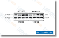 Click to enlarge | Rating | |
| Source | EMBO Mol Med, 2013, 5, 397–412. MG-132 purchased from Selleck | |
| Method | Western Blot | |
| Cell Lines | MEF cells | |
| Concentrations | 30 uM | |
| Incubation Time | 6 h | |
| Results | Treatment of MEF with proteasome inhibitor MG132 resulted in similar increase of steady state levels of intracellular ATZ in both GFP- and TFEB- transfected Cells. |
MiR-138 downregulates miRNA processing in HeLa cells by targeting RMND5A and decreasing Exportin-5 stability. [Li J, et al. Nucleic Acids Res 2014;42(1):458-74]
Gene transfer of master autophagy regulator TFEB results in clearance of toxic protein and correction of hepatic disease in alpha-1-anti-trypsin deficiency. [Pastore N, et al. EMBO Mol Med 2013;5(3):397-412]
Azacytidine and erlotinib exert synergistic effects against acute myeloid leukemia. [Lainey E, et al. Oncogene 2012;32(37):4331-42]