
Antibodies


Hesperadin potently inhibits Aurora B with IC50 of 250 nM. It markedly reduces the activity of AMPK, Lck, MKK1, MAPKAP-K1, CHK1 and PHK while it does not inhibit MKK1 activity in vivo.
| Size | Price | Stock | Quantity | ||||||||||||||||||||
|---|---|---|---|---|---|---|---|---|---|---|---|---|---|---|---|---|---|---|---|---|---|---|---|
| |||||||||||||||||||||||
 Distributor Tel: +886-2-2827-1197jimmy@new.abscience.com.tw
| Description | Hesperadin potently inhibits Aurora B with IC50 of 250 nM. It markedly reduces the activity of AMPK, Lck, MKK1, MAPKAP-K1, CHK1 and PHK while it does not inhibit MKK1 activity in vivo. | |||||
|---|---|---|---|---|---|---|
| Targets | Aurora B (human) | TbAUK1 | ||||
| IC50 | 250 nM [1] | 40 nM [2] | ||||
| In vitro | Hesperadin inhibits the ability of immunoprecipitated Aurora B to phosphorylate histone H3 with IC50 of 250 nM and markedly reduces the activity of other kinases (AMPK, Lck, MKK1, MAPKAP-K1, CHK1, and PHK) at a concentration of 1 μM. In contrast, only 20-100 nM of Hesperadin is sufficient to induce the loss of mitotic histone H3-Ser10 phosphorylation in HeLa cells. Hesperadin treatment causes defects in mitosis and cytokinesis, leading to stoppage of proliferation of HeLa cells and polyploidization, which can be specifically ascribed to the inhibition of Aurora B function during the process of chromosome attachment. Hesperadin (100 nM) quickly overrides the mitotic arrest induced by taxol or monastrol but not by nocodazole. Hesperadin and nocodazole treatment in HeLa cells abolishes kinetochore localization of BubR1 and diminishes the intensity of Bub1 at kinetochores, suggesting that Aurora B function is required for efficient kinetochore recruitment of BubR1 and Bub1, which in turn might be necessary for prolonged checkpoint signaling. [1] Hesperadin prevents the phosphorylation of recombinant trypanosome histone H3 by the T. brucei Aurora kinase-1 (TbAUK1) from pathogenic Trypanosoma brucei with IC50 of 40 nM in vitro kinase assays. Hesperadin significantly inhibits cell growth of cultured infectious bloodstream forms (BF) with IC50 of 48 nM, and only weakly inhibits cell growth of insect stage procyclic forms (PF) with IC50 of 550 nM. [2] | |||||
| In vivo | ||||||
| Features | ||||||
| The Aurora B kinase assay | For the Aurora B kinase assay, HeLa cells are lysed in a buffer containing 50 mM NaCl. The whole cell extract is spun at 13,000 rpm for 20 minutes at 4 °C using a table top centrifuge. The pellet obtained from 200 mg of whole cell extract is extracted again in 15 mL lysis buffer containing 250 mM NaCl in order to obtain active Aurora B kinase from mitotic chromatin. The low speed supernatant of the latter extract is used for immunoprecipitation. Monoclonal mouse anti–AIM-1, or mouse anti-HA, is coupled to GammaBind Plus Sepharose, and beads are rotated over-end in the extract for 90 minutes at 4 °C. Beads are washed, aliquoted, and washed in kinase buffer (20 mM Tris, pH 7.5, 150 mM NaCl, 10 mM MgCl2, 1 mM DTT, 10 mM NaF). The kinase assay is performed with 10 μL beads in 20 μL kinase buffer containing 5 μg histone H3, 10 μM ATP, 2.5 μCi [γ-32P]ATP, and different concentrations of Hesperadin for 20 minutes at 37 °C. SDS sample buffer is added, and samples are boiled and resolved by SDS-PAGE. The gel is dried, and the radioactive signal is detected by PhosphorImager analysis. The data is analyzed using ImageQuant software. |
|---|
| Cell lines | HeLa cells and PtK1 cells |
|---|---|
| Concentrations | Final concentration ~500 nM |
| Incubation Time | 24 and 48 hours |
| Method | Cells are exposed to different concentrations of Hesperadin for 24 and 48 hours. At indicated time points, methanol-fixed cell samples are washed with PBS and subsequently stained in PI buffer (50 μg/mL propidium iodide, 10 mM Tris, pH 7.5, 5 mM MgCl2, 200 μg/mL RNase A) for 20-40 minutes at 37 °C. The DNA content is determined by flow cytometry. |
| Molecular Weight (MW) | 516.65 |
|---|---|
| Formula | C29H32N4O3S |
| CAS No. | 422513-13-1 |
| Storage | 3 years -20℃Powder |
|---|---|
| 6 months-80℃in DMSO | |
| Syonnyms | N/A |
| Solubility (25°C) * | In vitro | DMSO | 103 mg/mL (199 mM) |
|---|---|---|---|
| Water | <1 mg/mL (<1 mM) | ||
| Ethanol | <1 mg/mL (<1 mM) | ||
| In vivo | 30% PEG400/0.5% Tween80/5% propylene glycol, | 30 mg/mL | |
| * <1 mg/ml means slightly soluble or insoluble. * Please note that Selleck tests the solubility of all compounds in-house, and the actual solubility may differ slightly from published values. This is normal and is due to slight batch-to-batch variations. | |||
| Chemical Name | (Z)-N-(2-oxo-3-(phenyl(4-(piperidin-1-ylmethyl)phenylamino)methylene)indolin-5-yl)ethanesulfonamide |
|---|
| Stock Solution (1ml DMSO) | 1mM | 10mM | 20mM | 30mM |
|---|---|---|---|---|
| Mass(mg) | 0.51665 | 5.1665 | 10.333 | 15.4995 |
 Click to enlarge | Rating | |
| Source | Hesperadin purchased from Selleck | |
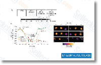
| Method | Cdk1 reactivation assay | |
| Cell Lines | U2OS cell | |
| Concentrations | 125 nM | |
| Incubation Time | 25 min | |
| Results |
Haspin inhibitors reveal centromeric functions of Aurora B in chromosome segregation. [Wang F, et al. J Cell Biol 2012;199(2):251-68]
Aurora B potentiates Mps1 activation to ensure rapid checkpoint establishment at the onset of mitosis. [Saurin AT, et al. Nat Commun 2011;2:316]
A complex of Kif18b and MCAK promotes microtubule depolymerization and is negatively regulated by Aurora kinases. [Tanenbaum ME, et al. Curr Biol 2011;21(16):1356-65]