
Antibodies


Canertinib (CI-1033) is a pan-ErbB inhibitor for EGFR and ErbB2 with IC50 of 1.5 nM and 9.0 nM, no activity to PDGFR, FGFR, InsR, PKC, or CDK1/2/4. Phase 3.
| Size | Price | Stock | Quantity | ||||||||||||||||
|---|---|---|---|---|---|---|---|---|---|---|---|---|---|---|---|---|---|---|---|
| |||||||||||||||||||
 Distributor Tel: +886-2-2827-1197jimmy@new.abscience.com.tw
| Description | Canertinib (CI-1033) is a pan-ErbB inhibitor for EGFR and ErbB2 with IC50 of 1.5 nM and 9.0 nM, no activity to PDGFR, FGFR, InsR, PKC, or CDK1/2/4. Phase 3. | |||||
|---|---|---|---|---|---|---|
| Targets | EGFR | ErbB2 | ||||
| IC50 | 1.5 nM | 9.0 nM [1] | ||||
| In vitro | CI-1033 shows excellent potency for irreversible inhibition of erbB2 autophosphorylation in MDA-MB 453 cells. CI-1033 also shows high permeability in Caco-2 cells and inhibits secretory transport of vinblastine, which indicates that CI-1033 is a likely inhibitor of the P-gp. [1] CI-1033 alone, significantly suppresses constitutively activated Akt and MAP kinase. In combination with gemcitabine, CI-1033 inhibits Akt and prevents increased levels of MAPK phosphorylation. CI-1033 stimulates p27 expression and p38 phosphorylation in MDA-MB-453 cells. [2] CI-1033 is highly specific to the erbB receptor family and not sensitive to PGFR, FGFR or IR even at 50 μM. CI-1033 shows high levels of inhibition in A431 cells expressing EGFR with IC50 of 7.4 nM. CI-1033 suppresses heregulin-stimulated tyrosine phosphorylation of erbB2, erbB3 and erbB4 with IC50 of 5, 14 and 10 nM, respectively. CI-1033 also inhibits expression of pp62c-fos in response to heregulin. [3] CI-1033 is predicted to modify Cys773 covalently within the ATP binding site of the HER2 kinase and enhances destruction of both mature and immature ErbB-2 molecules. [4] CI-1033 induces a significant decrease in measurable phosphorylation of tyrosine residues 845 and 1068 of EGFR, which are responsible for Src and Ras/MAPK signaling respectively. The corresponding residues of Her-2, tyrosine residues 877 and 1248 are dephosphorylated significantly by CI-1033 at a concentration of 3 μM or higher. CI could block EGFR internalization and increase the rate of apoptosis in primary osteosarcoma cells in a titratable fashion. [5] In addition, CI-1033 inhibits the proliferation of TT, TE2, TE6 and TE10 cells significantly at 0.1 nM. [6] | |||||
| In vivo | CI-1033 shows impressive activity against A431 xenografts in nude mice at 5 mg/kg of body weight. [1] CI-1033 (20 to 80 mg/kg/d) achieves a high degree of tumor regressions in H125 xenograft models. [3] Oral administration of CI-1033 causes a marked inhibition of growth in TT, TE6 and TE10 xenografts in nude mice, without animal death and <10% weight loss. [6] | |||||
| Features | First kinase inhibitor to show irreversible activity and to have entered clinical trials (serving as a template for further development). | |||||
| Tyrosine Kinase Assays | Enzyme assays for determination of IC50 are performed in 96-well filter plates in a total volume of 0.1 mL, containing 20 mM Hepes, pH 7.4, 50 mM sodium vanadate, 40 mM magnesium chloride, 10 μM adenosine triphosphate (ATP) containing 0.5 mCi of [32P]ATP, 20 mg of polyglutamic acid/tyrosine, 10 ng of EGFR tyrosine kinase, and appropriate dilutions of CI-1033. All components except the ATP are added to the well and the plate is incubated with shaking for 10 min at 25 °C. The reaction is started by adding [32P]ATP, and the plate is incubated at 25 °C for another 10 min. The reaction is terminated by addition of 0.1 mL of 20% trichloroacetic acid (TCA). The plate is kept at 4 °C for at least 15 min to allow the substrate to precipitate. The wells are then washed five times with 0.2 mL of 10% TCA and 32P incorporation determined with a Wallac β plate counter. |
|---|
| Cell lines | TT, TE2, TE6 and TE10 cells |
|---|---|
| Concentrations | 0.1-5.0 nM |
| Incubation Time | 1, 3, 5 and 7 days |
| Method | Cells (1 × 10⁴) are seeded in each well of a 24-well plastic culture plate and left overnight in DMEM or RPMI-1640 supplemented with 10% FBS. The next morning, the cells are treated with the indicated concentrations of CI-1033 (0.1-5.0 nM) for varying periods (1, 3, 5 and 7 days). After treatment, the cells are counted using a Coulter counter. The percent of cell proliferation is calculated by this formula: treatment cell number/control cell number × 100 for each time period. |
| Animal Models | A431 xenografts established in nude mice |
|---|---|
| Dosages | ~18 mg/kg |
| Administration | Administered orally |
| Solubility | 30% propylene glycol, 5% Tween 80, 65% D5W, 10 mg/mL |
| NCT Number | Recruitment | Conditions | Sponsor /Collaborators | Start Date | Phases |
|---|---|---|---|---|---|
| NCT00051051 | Completed | Breast Neoplasms | Pfizer | 2002-12 | Phase 2 |
| NCT00174356 | Completed | Carcinoma, Non-Small Cell Lung | Pfizer | 2002-12 | Phase 1 |
| NCT00050830 | Completed | Lung Neoplasms | Pfizer | 2003-01 | Phase 2 |
| NCT00362986 | Completed | Dermatologic Complications|Unspecified Adult Solid Tumor, Protocol Specific | National Cancer Institute (NCI)|North Central Cancer Treatment Group | 2006-10 | Phase 3 |
Download Canertinib (CI-1033) SDF
| Molecular Weight (MW) | 485.94 |
|---|---|
| Formula | C24H25ClFN5O3 |
| CAS No. | 267243-28-7 |
| Storage | 3 years -20℃Powder |
|---|---|
| 6 months-80℃in DMSO | |
| Syonnyms | PD-183805 |
| Solubility (25°C) * | In vitro | DMSO | 2 mg/mL (4 mM) |
|---|---|---|---|
| Water | <1 mg/mL (<1 mM) | ||
| Ethanol | 9 mg/mL (18 mM) | ||
| In vivo | 30% propylene glycol, 5% Tween 80, 65% D5W, | 10 mg/mL | |
| * <1 mg/ml means slightly soluble or insoluble. * Please note that Selleck tests the solubility of all compounds in-house, and the actual solubility may differ slightly from published values. This is normal and is due to slight batch-to-batch variations. | |||
| Chemical Name | N-(4-(3-chloro-4-fluorophenylamino)-7-(3-morpholinopropoxy)quinazolin-6-yl)acrylamide |
|---|
 Click to enlarge | Rating | |
| Source | Cell Cycle 8, 13, 2050-2056. Canertinib (CI-1033) purchased from Selleck | |
| Method | Western Blot | |
| Cell Lines | H1993 cells | |
| Concentrations | 1.5 uM | |
| Incubation Time | 4 h | |
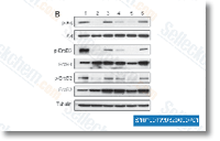
| Results | We pretreated the cells with SU11274 for 48 h before adding, for the final 4 h, the PKC δ inhibitor rottlerin, GO6976 (an inhibitor blocking the activities of classical PKCs only), or CI-1033 (an irreversible ErbB TKI). Inclusion of rottlerin for the final 4 h effectively inhibited the re-activation of Akt, ErbB2 and ErbB3 (Fig. 4B). GO6976 was ineffective, while CI-1033 had an intermediate impact on the restoration of ErbB/AKT signaling. |
Differential sensitivity of glioma- versus lung cancer-specific EGFR mutations to EGFR kinase inhibitors. [Vivanco I, et al. Cancer Discov 2012;2(5):458-71]
The fibroblast-derived paracrine factor neuregulin-1 has a novel role in regulating the constitutive color and melanocyte function in human skin. [Choi W, et al. J Cell Sci 2010;123(Pt 18):3102-11]
Secretory phospholipase A2-IIa is involved in prostate cancer progression and may potentially serve as a biomarker for prostate cancer. [Dong Z, et al. Carcinogenesis 2010;31(11):1948-55]