
Antibodies


AG-1024 inhibits IGF-1R autophosphorylation with IC50 of 7 μM, is less potent to IR with IC50 of 57 μM and specifically distinguishes between InsR and IGF-1R (as compared to other tyrphostins).
| Size | Price | Stock | Quantity | ||||||||||||
|---|---|---|---|---|---|---|---|---|---|---|---|---|---|---|---|
| |||||||||||||||
 Distributor Tel: +886-2-2827-1197jimmy@new.abscience.com.tw
| Description | AG-1024 inhibits IGF-1R autophosphorylation with IC50 of 7 μM, is less potent to IR with IC50 of 57 μM and specifically distinguishes between InsR and IGF-1R (as compared to other tyrphostins). | |||||
|---|---|---|---|---|---|---|
| Targets | IGF-1R | IR | ||||
| IC50 | 7 μM | 57 μM [1] | ||||
| In vitro | AG-1024 blocks the IGF-1 receptor and IR autophosphorylation with IC50 of 7 μM and 57 μM, respectively. AG-1024 also inhibits the receptor tyrosine kinase activity towards exogenous substrates (TKA) with IC50 values of 18 μM and 80 μM, respectively. [1] AG-1024 (10 μM) inhibits cell proliferation in a time-dependent manner, and induces apoptosis in MCF-7 cells at 48 hours by 20.1% and >40% when combined with irradiation (10 Gy), more potently than that of irradiation (10 Gy) alone by 11.8%, which is associated with a down-regulation of phospho-Akt1 and bcl-2, and up-regulation of Bax, p53 and p21. [2] AG-1024 significantly inhibits melanoma cell proliferation with an IC50 of <50 nM in the absence of serum, by blocking MAPK/ERK2 signaling, subsequently rapidly inducing pRb dephosphorylation and activation, and eventually the formation of growth suppressive pRb-E2F complexes. [3] AG-1024 treatment down-regulates the expression of Bcr-Abl and P-Akt, and up-regulates DNA-PKcs expression in UT7-9 and Ba/F3-p210 cells, leading to decreased clonogenic survival and proliferation. AG-1024 also significantly inhibits the proliferation of cells resistant to the BCR-ABL inhibitor STI571, which correlates with a dose-dependent decrease in Bcr-Abl protein expression. [4] | |||||
| In vivo | Administration of AG-1024 at a dose of 30 μg for 10 days significantly inhibits the tumor growth of Ba/F3-p210 xenograft in mice. [4] | |||||
| Features | ||||||
| Inhibition of IGF-1- and insulin-stimulated cellular proliferation | NIH-3T3 cells overexpressing IGF-1 or insulin receptors are plated on 96-well plates (2,000-5,000 cells/well) and maintained overnight in complete medium. Cells are then changed to DMEM containing 1% FBS in the presence of 10 nM IGF-1 or insulin and various concentrations of AG-1024 for 120 hours. Medium is replaced every 48 hours. At the indicated periods of time, the medium is aspirated from the wells and 100 μL MTT is added to each well. The cells are then incubated for 4 hours at 37 °C and lysed by addition of 100 μL isoamylic alcohol and shaking for 20 minutes. The plate is then read in the ELISA reader at 570 and 690 nm. The IC50 value is calculated at the 120-hour time point. |
|---|
| Cell lines | MCF-7 |
|---|---|
| Concentrations | Dissolved in DMSO, final concentration 10 μM |
| Incubation Time | 24, 48 or 72 hours |
| Method | Cells are exposed to AG-1024 for 24, 48 or 72 hours. For the determination of proliferation, cells are harvested and counted with trypan blue dye exclusion. Apoptosis is evaluated by dual staining of MCF-7 with fluoresceine anti-digoxigenin and propidium iodide. Briefly, fixed cells are washed with PBS, suspended in TdT buffer with TdT enzyme and Dig-dUTP for 60 minutes, and suspended in FITC blocking solution with anti-Dig-Fluorescein for 30 minutes at room temperature and kept in a dark place. Cells are then rinsed in buffer and resuspended in propidium iodide/RNase A solution for 30 minutes then analyzed by flow cytometry. For the assessment of phospho-Akt1, Bax, p53, bcl-2 and p21, cells are lysed and analyzed by western blot. |
| Animal Models | Female nude mice implanted subcutaneously with Ba/F3-p210 cells |
|---|---|
| Dosages | 30 μg/day |
| Administration | Injected i.p |
| Solubility | 1% DMSO/30% polyethylene glycol/1% Tween 80, 30 mg/mL |
| Molecular Weight (MW) | 305.17 |
|---|---|
| Formula | C14H13BrN2O |
| CAS No. | 65678-07-1 |
| Storage | 3 years -20℃Powder |
|---|---|
| 6 months-80℃in DMSO | |
| Syonnyms | N/A |
| Solubility (25°C) * | In vitro | DMSO | 61 mg/mL (199 mM) |
|---|---|---|---|
| Water | <1 mg/mL (<1 mM) | ||
| Ethanol | <1 mg/mL (<1 mM) | ||
| In vivo | 1% DMSO/30% polyethylene glycol/1% Tween 80, | 30 mg/mL | |
| * <1 mg/ml means slightly soluble or insoluble. * Please note that Selleck tests the solubility of all compounds in-house, and the actual solubility may differ slightly from published values. This is normal and is due to slight batch-to-batch variations. | |||
| Chemical Name | 2-(3-bromo-5-tert-butyl-4-hydroxybenzylidene)malononitrile |
|---|
 Click to enlarge | Rating | |
| Source | Mol Endocrinol, 2011, 25(12), 2041-53. AG-1024 purchased from Selleck | |
| Method | Cell viability assay | |
| Cell Lines | A549 cells | |
| Concentrations | 0-10 μM | |
| Incubation Time | 48 h | |
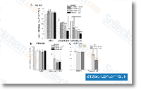
| Results | Treating A549 cells with AZD6244 alone at concentrations rang-ing from 100 nM to 5 μMhad no effect on cell viabilities (Fig. A). However, when combined with 1 and 2 μM gossypol, AZD6244 noticeably reduced the number of viable cells in a dose-dependent manner. We also examined the inhibitory effects of an EGFR tyrosine kinase inhibitor AG1478 and an IGF-I receptor (IGF-IR) inhibitor AG1024 on breast cancer cell viability in combination with gossypol. Exposure to a low concentration of gossypol significantly sensitized SKBR3 and MCF-7 breast cancer cells to AG1478 and AG1024, respectively (Fig. B and C) |
Insulin-like growth factor 1 receptor (IGF1R) as a novel target in chronic lymphocytic leukemia. [Yaktapour N, et al. Blood 2013;122(9):1621-33]